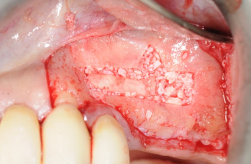
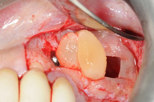
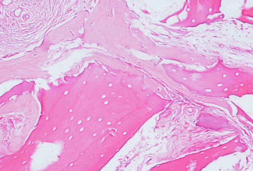
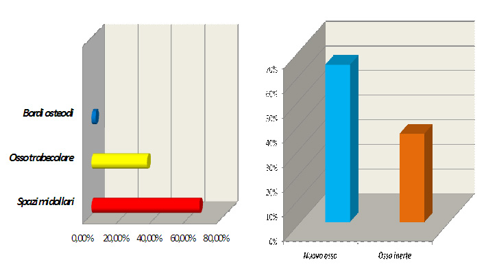
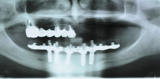
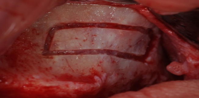
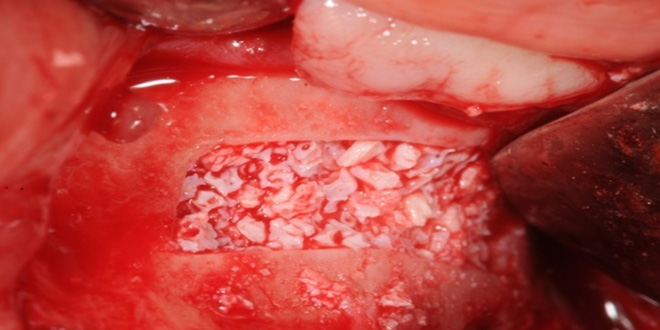
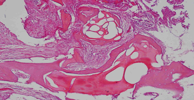
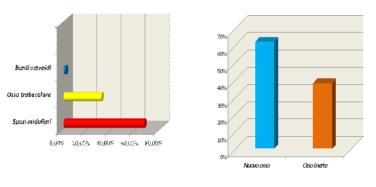
The aim of the present study is to assess, through clinical and histological evaluations, the regenerative potential of PRF® in association with heterologous bone grafts (Bio-Oss®) for the treatment of maxillary atrophies using the sinus-lift procedure, in comparison with the use of heterologous graft only.

MATERIALS AND METHODS
This study was carried out on a group of 15 patients presenting with posterior maxillary atrophy with residual crest < 5mm and requiring implant-prosthetic rehabilitation. Two different groups were formed according to the grafting material used: the Test Group received PRF/Bio-Oss® for a total of 11 sinus lifts, and the Control Group received only Bio-Oss® as grafting-material for a total of 6 sinus lifts.

The implants were placed 4 months later for the Test Group and 8 months later for the Control Group, and at the same time, trephine biopsy was performed for histological evaluation.

RESULTS
In the Test Group, the histological results showed the presence of lamellar bone tissue with an intensely-eosinophilic bone matrix mixed with fragments of lamellar bone tissue with inhabited osteocyte lacunae and a slightly-eosinophilic bone matrix, probably to be ascribed to the newly-formed bone tissue; in the Control Group, 8 months later we can observe optically-empty globular lacunae, a result of the Bio-Oss granules not yet integrated into the bone matrix. Histomorphometric analysis showed a substantial similarity between the rate of trabecular bone and medullary spaces in the two groups.

Test group

Control group

  TEST GROUP CONTROL GROUP
Innesto osseo PRF/Bio-Oss   Bio-Oss
Tempo di prelievo Dopo 4 mesi   Dopo 8 mesi
N° sinus-lift    11    6
Spazi midollari%    64,5    66,7
Bordi osteoidi%    2,26    1,94
Osso trabecolare%    33,24    31,36
osso inerte% (fraz trab)    36    38
osso vitale % (fraz trab)    64    62

DISCUSSION
Only 4 months later, the use of PRF® seems to have produced good neoangiogenesis, reducing the areas of non-vital bone if compared to the Control Group, even though in statistically insignificant quantity due to the small sample.

CONCLUSIONS
Being rich in fibrin, platelets, leukocytes and growth factors, PRF® resulted in a bone regeneration which allowed to place the fixtures already 4 months after surgery. Therefore, as the histological and histomorphometric analysis revealed, PRF® reduces the time needed for bone healing, in comparison with the only use of Bio-Oss®.

Università degli Studi di Bari “A. Moro” Dipartimento di Odontostomatologia e Chirurgia
XX Congresso nazionale Docenti di Odontoiatria

Autori:
F. Inchingolo, A. Colella , M. Marrelli, A. Palladino, M. Tatullo, A.M. Inchingolo, F. Carbotti, A.D. Inchingolo, G. Malcangi, F. Schinco, M. Serafini, G. Marinelli, G. Dipalma

BIBLIOGRAFIA
Dohan Ehrenfest DM, Del Corso M, Diss A, Mouhyi J, Charrier JB. Three-dimensional architecture and cell compositions of a Choukroun’s platelet-rich fibrin clot and membrane. J Periodontol 2010;81:546-55 Yilderman M., Spiekermann H., Handt., Edelhoff D. Maxillary sinus augmentation with tha xenograft Bio-Oss and autogenous intraoral bone for qualitative improvement of the implant site: a histologic and histomorphometric clinical study in humans. Misch CE, Judy KWM. “Classification of partially edentulous arches for implant dentistry”. Int J Oral Implant 1987; 4: 7-12.

Lascia un commento

Il tuo indirizzo email non sarà pubblicato. I campi obbligatori sono contrassegnati *