White Sponge Nevus (WSN) is a rare pathology with a pathogenesis on genetic basis, a benign course and a localization affecting the mucosal keratin.
WSN is usually a symptomless pathology: when pain is present, some authors reported re-duction of symptoms by taking penicillin or oral tetracycline rinses, suggesting that a bacterial overinfection could be at the base of possible painful symptoms.
Case Report. We describe 2 patients affected by WSN, father and son: they presented two different oral diseases associated with an infection by Staphylococcus aureus. So, we have performed a careful oral hygiene to reduce infection in the oral cavity. In the following days we prescribed 2 rinses a day with a mouthwash containing chlorhexidine digluconate at two different percentages.
Discussion. Early diagnosis of this lesion is important, because it allows us to exclude other more serious diseases. In the most part of cases, WSN requires no treatment because of its benign and asymptomatic behaviour: up to now, no protocol of treatment for this condition was standardized. Even if WSN is a painless condition, sometime a correlated painful symptomatology was reported.
Conclusions. In our experience, we have achieved excellent results even with chlorhexidine digluconate rinses, considering that our treated cases were both infected by Staphylococcus aureus.
We hypothesize that the corrugated plaques and the altered texture of the mucosa create the right conditions for the colonization and the development of microbial species such as sap-rophytic bacteria or fungal species.
Case Report
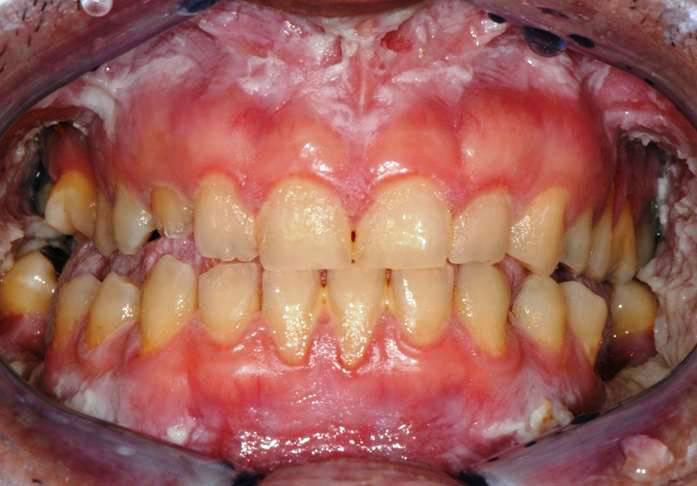
An 38 year-old Italian man came to the Depart-ment of Oral Hygiene and Periodontology at Calabrodental clinic in April 2011 for the evaluation of white bilateral corrugated plaques developed on the buccal mucosa and on the gingiva, these plaques were been present since he was 13 years-old. (Fig.1)
This clinical condition was investigated by an-other clinician who carried out a diagnosis of White Sponge Nevus, excluding so the other similar pa-thologies compatible with this clinical aspect.
These plaques were always asymptomatic, however, from 2 months, the patient complained of burning symptoms that persisted throughout the day. So, he came at the clinic Calabrodental in order to alleviate these symptoms. After routine investiga-tions, we prescribed an oral buffer to assess the pos-sible presence of bacterial or fungal superinfection: the result was positive for Staphylococcus aureus which showed a susceptibility to methicillin. The analysis didn’t evidenced the presence of Candida albicans or other fungal infections, so the painful symptomatology could be related to a poor oral hy-giene and to the presence of the methicillin-resistant Staphylococcus aureus in the oral cavity of our pa-tient.
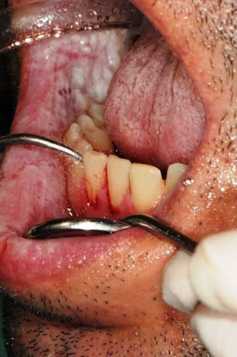
So, we have performed a careful oral hygiene to reduce infection in the oral cavity. (Fig.2)
In the following days we prescribed 2 rinses a day with a mouthwash containing chlorhexidine di-gluconate at 0.2%. We performed the follow-up after 7 days and the patient has reported the disappearance of pain. During the check we noted that the same plaques were developed on the oral mucosa of the son of our patient, so we wanted to document the case, after obtaining the written consent of the patient.
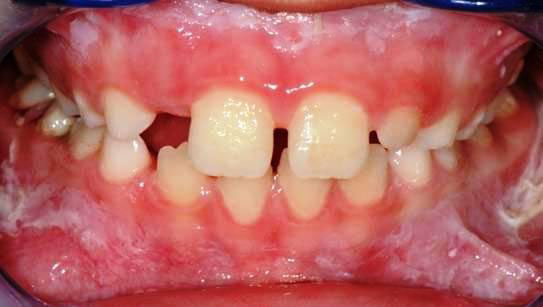
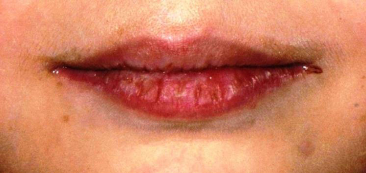
Our little patient showed an intraoral clinical condition very similar to father’s, moreover, the pa-tient was affected by a bilateral angular cheilitis. (Fig.3,4).
A buffer containing patient’s saliva was submit-ted to a diagnostic oral microbiology laboratory: the microbiological analysis showed in the presence of Staphylococcus aureus sensitive to methicillin (MSSA) in the oral cavity of our young patient, while, no fungal infections was found.
2 rinses a day with mouthwash containing chlorhexidine digluconate at 0,12% was prescribed in order to decrease the bacteria and the occurrence of relapses of angular cheilitis.
Discussion
The aspect of WSN is not strongly pathogno-monic. It’s important to perform careful clinical and histological examinations to differentiate this benign condition from other potentially pre-malignant le-sions as well as oral lichen planus, keratosis follicu-laris, candidiasis, lichenoid reactions and lupus ery-thematosus12.
Early diagnosis of this lesion is important, be-cause it allows us to exclude other more serious dis-eases. In the most part of cases, WSN requires no treatment because of its benign and asymptomatic behaviour: up to now, no protocol of treatment for this condition was standardized 13. Even if WSN is a painless condition, sometime a correlated painful symptomatology was reported 5,10,11.
Antibiotic treatment with oral penicillin 10, am-picillin 5,11, and tetracycline has achieved a moderate success; some authors have also suggested the use of tetracycline mouthwashes. In our experience, we have achieved excellent results even with chlorhexidine digluconate rinses, considering that our treated cases were both infected by Staphylococcus aureus. WSN is not considered a bacterial disease; however, since antibiotic therapy is able to reduce the symptomatol-ogy occasionally occurred, it is possible that infections may play a role in the pathogenesis of this disease 14. Furthermore, the corrugated plaques and the altered texture of the mucosa create the right conditions for the colonization and the development of microbial species such as saprophytic bacteria or fungal species. In these 2 reported cases, the wide part of mucosa interested by the plaques of the WSN is a condition which promotes the grown of the Staphylococcus aure-us 15.
Conclusions
In all reported cases, the patients affected by WSN achieve a condition of clinical stability between 20 and 30 years, moreover, have never been described any malignant transformation of the interested mu-cosa16; however, if a Staphylococcus aureus infection is developing, there may be periods of exacerbation and remission of the symptoms: so we suggest to take care of the oral hygiene and to daily use a mouthwash containing chlorhexidine 0.05% in order to prevent the growth of most dangerous species of bacteria, especially the Staphylococcus aureus as we reported in our communication.
Authors’ Contributions
MM and SB participated in the clinical and pharmacological management of the treated cases.
MT drafted the manuscript and revised the lit-erature sources.
FI participated in the follow-up examinations.
All authors read and approved the final manu-script.
Consent Statement
Written informed consent was obtained from the patient for publication of this case report and accom-panying images. A copy of the written consent is available for review by the Editor-in-Chief of this journal.
Conflict of Interest
All authors disclose any financial and personal relationships with other people or organizations that could inappropriately influence (bias) their work.
References
1. Cannon AB. White sponge nevus of the mucosa (naevus spon-giosus albus mucosae). Arch Dermatol Syphilol 1935; 31: 365
2. Woo S-B. Diseases of the oral mucosa. In: Mckee PH, et al., eds. Pathology of the Skin with Clinical Correlations. 3rd ed. Phila-delphia: Elsevier Mosby, 2005:387
3. Frithiof L, Banoczy J. White sponge nevus (leukoedema exfoli-ativum mucosae oris): ultrastructural observations. Oral Surg 1976; 41: 607
4. Jorgensen RJ, Levin S. White sponge nevus. Arch Dermatol 1981; 117: 73
5. Sadeghi EM, Witkop CJ. The presence of Candida albicans in hereditary benign intraepithelial dyskeratosis: an ultrastruc-tural observation. Oral Surg Oral Med Oral Pathol 1979; 48:342-346.
6. Jorgenson RJ, Levin LS. White sponge nevus. Arch Dermatol 1981; 117:73-76.
7. Yavazyilmaz E, et al. Oral-dental findings in dyskeratosis con-genita. J Oral Pathol Med 1992; 21:280-284.
8. Martelli Jr H, Pereira SM, Rocha TM, Santos PLAN, Paula AMB, Bonan RF. White sponge nevus: report of a three-generation family. Oral Surg Oral Med Oral Pathol Oral Radiol Endod 2007; 103:43-7.
9. López Jornet P. White sponge nevus: presentation of a new family. Pediatr Dermatol. 2008;25(1):116-7
10. Alinovi A, Benoldi D, Pezzarossa E. White sponge nevus: suc-cessful treatment with penicillin. Acta Derm Venerol 1983;63:83-5
11. Lim J, Ng SK. Oral tetracycline rinse improves symptoms of white sponge nevus. J Am Acad Dermatol 1992;26:1003-5.
12. Inchingolo F, Tatullo M, Abenavoli FM, Marrelli M, Inchingolo AD, Inchingolo AM, Dipalma G. Non-Hodgkin lymphoma af-
fecting the tongue: unusual intra-oral location. Head Neck Oncol. 2011 Jan 4;3:1.
13. Sambucety OS, López PM, Prieto MAR, Gónzalez IR, Fernán-dez MM. Lesiones blanquecinas en la mucosa oral. An Esp Pe-diatr 2001; 55:159-60
14. McDonagh AJG, et al. White sponge naevus successfully treated with topical tetracycline. Clin Exp Dermatol 1990;15:152
15. Salgodo CO, Farr BM, Calfee DP. Community-acquired methi-cillin-resistant Staphylococcus aureus: a meta-analysis of prev-alence and risk factors. Clin Infect Dis 2003; 36: 131–9
16. Lamey PJ, et al. Oral White sponge naevus: response to antibi-otic therapy. Clin Exp Dermatol 1998;23:59
 |
 |
 |
| Fig. 1 | Fig. 2 | Fig. 3 |
 |
||
| Fig. 4 | ||
Keywords: Chirurgia orale; Cisti solitaria; Pseudocisti; Cisti traumatica; Platelet-rich fibrin Keywords Oral surgery; Solitary cyst; Pseudocyst; Traumatic cyst; Platelet-rich fibrin
Autori
M. Marrelli, M. Tatullo, G. Di Palma, F. Inchingolo