The aim of the present study is to define Eagle syndrome, a rare and underestimated condition, and to include it in a differential diagnosis with other conditions presenting similar signs and symptoms.
The literature describes Eagle syndrome as the symptomatic elongation of the styloid process and the calcification of the stylohyoid ligament. It involves a series of painful, sometimes disabling symptoms to craniofacial and cervical regions, such as dysphagia, odynophagia, facial pain, otalgia, headache and trismus.
Two manifestations of this syndrome can be distinguished: the first manifestation affects tonsillectomized patients and is characterized by pharyngeal pain irradiating to the middle ear and mastoid region; the second form causes spontaneous pain in distribution territories of the internal and/or external carotid and possible faintness or lipothymia with head rotation because of the temporary occlusion of the internal carotid.
MATHERIALS AND METHODS
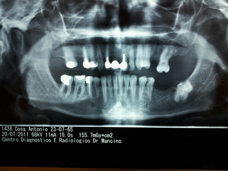
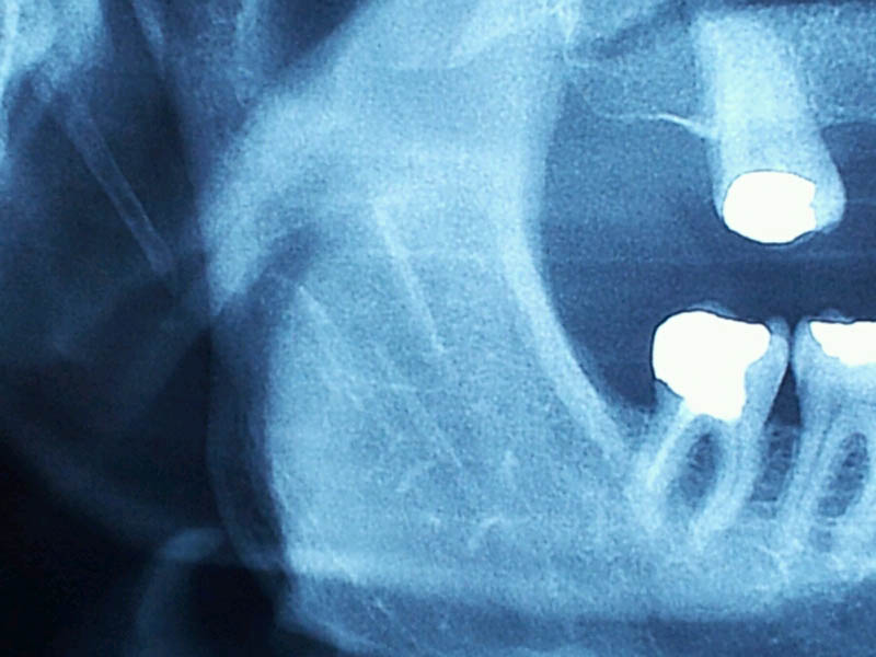
The diagnosis is based on clinical and radiographic examinations. These signs and symptoms lead to the diagnostic suspect. Palpation of the tonsillar fossa causes an exacerbation of symptoms as confirmed by instrumental examinations: orthopantomography (OPT) associated to 3d CT scan.
The therapeutic approach is strictly related to the seriousness of symptoms and can consists of medical maneuvers (infiltrations of corticosteroids and local anesthetics into the apex of the styloid process) or surgery (partial resection of the styloid process, with an intraoral or extraoral approach). The prognosis is excellent.
CASE REPORT
We report the case of a 58 year-old male patient, who complained of frequent headache, dysphagia, odynophagia and sporadic lipothymia. Palpation of the tonsillar fossa caused an exacerbation of symptoms.
He had been treated for many years with numerous bite without finding any improvement in symptoms.
OPT showed calcification of both stylohyoid ligaments. The patient was treated with infiltrations of corticosteroids and local anesthetics into the apex of the styloid process.
CONCLUSION
It is fundamental to understand which is the best diagnostic probe and the most adequate treatment for these patients suffering for many years and often undergoing useless therapies.
|
|
|
| Gingival vestibular areas of both jaws | Upper and lower right fornix |
Authors’ Contributions
INCHINGOLO Francesco, MARRELLI Massimo, PALLADINO Antonio, TATULLO Marco, INCHINGOLO Angelo Michele, CARBOTTI Filippo, DIPALMA Gianna, ANGELINI Valentina, INCHINGOLO Alessio Danilo, SERAFINI Maurizio, MALCANGI Giuseppina, SCHINCO Fabio, MARANO Giuseppe, CHIARAVALLOTTI Ernesto
University of Bari Department of Odontostomatology and Surgery – Dept. Head: Prof. D. Devito Calabrodental S.r.l. Operative Unit of Maxillo-Facial Surgery Regione Calabria – Crotone
References
Trauma induced eagle syndrome.Koivumäki A, Marinescu-Gava M, Järnstedt J, Sándor GK, Wolff J.Int J Oral Maxillofac Surg. 2012 Mar;41(3):350-3.
Epub 2012 Jan 12 Stylohyoid syndrome: surgical approach.Valerio CS, Peyneau PD, de Sousa AC, Cardoso FO, de Oliveira DR, Taitson PF, Manzi FR.J Craniofac Surg. 2012 Mar;23(2):e138-40
Eagle’s syndrome in an orthodontic patient.Pithon MM.Am J Orthod Dentofacial Orthop. 2012 Jan;141(1):113-5